How Salivary Glands Function Biology Diagrams Aside from the sublingual gland, the sublingual space is also comprised of the deep portion of the submandibular gland, the submandibular duct, the lingual artery/vein, and the lingual branch of the mandibular division of the trigeminal nerve (CN V3).[1,2,3,4] The sublingual gland itself is composed of a major sublingual gland and 8-30 small
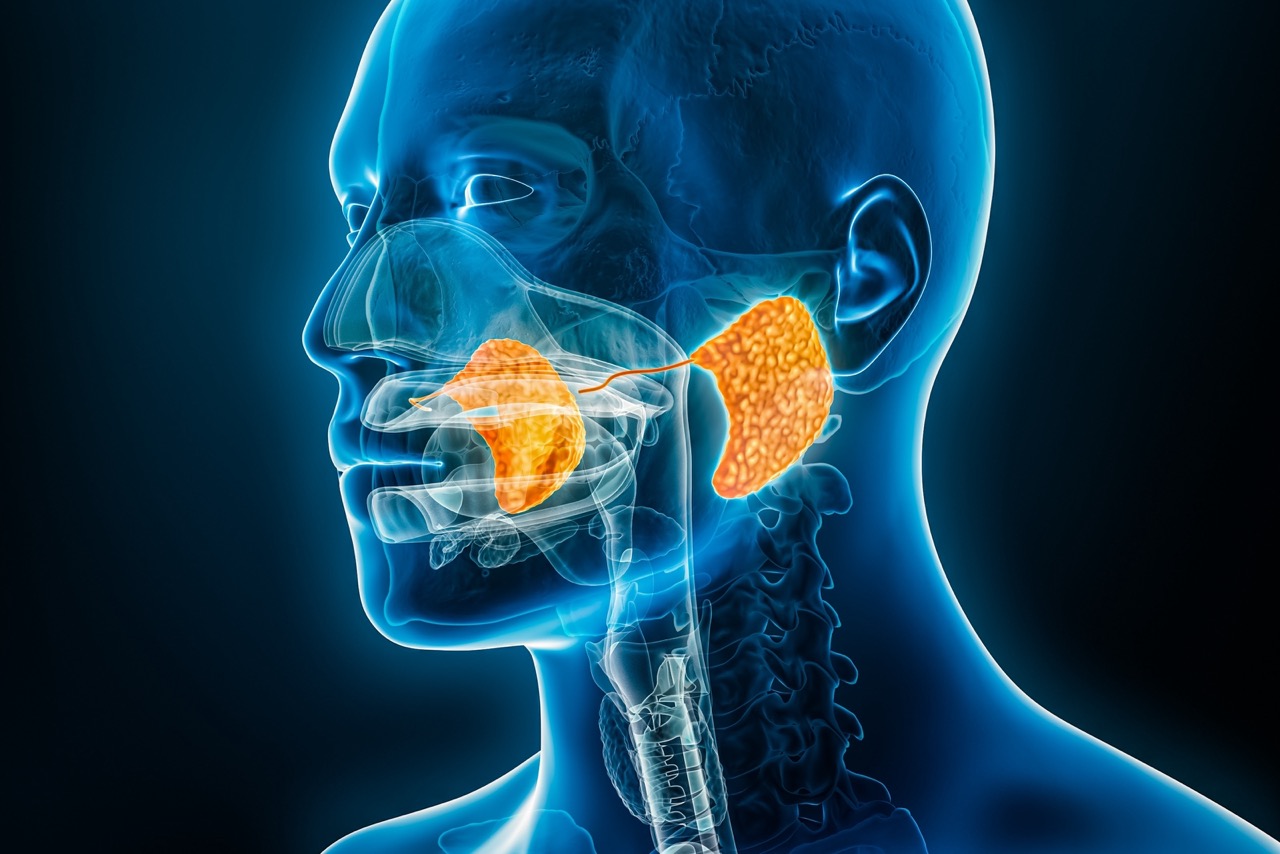
Your salivary glands lubricate your mouth, help you swallow, aid in digestion and help protect your teeth against harmful bacteria. You have three major types of salivary glands, including your sublingual, submandibular and parotid. Common symptoms of salivary gland disorders include fever, headaches and a lump in your cheek or under your chin. HIV (human immunodeficiency virus) and AIDS: The virus can cause salivary gland enlargement, fever, pain, and xerostomia (dry mouth).; Sjögren's disease: This autoimmune disease affects moisture-producing glands in the body and causes swelling and dry mouth; Diabetes: High blood glucose (sugar) may cause salivary gland enlargement and lower saliva production. Human salivary glands represent a crucial part of oropharyngeal anatomy. Saliva contains amylase, which initiates digestion through the breakdown of carbohydrates. Salivary gland pathology may be a harbinger of systemic disease, including conditions that may shorten the life span. These glands also play a crucial role in immunologic defense as

Review of the Major and Minor Salivary Glands, Part 1: Anatomy ... Biology Diagrams
Salivary glands are exocrine glands responsible for producing and secreting saliva into the oral cavity.[8] Saliva aids in moistening food, digestion, lubrication, and oral hygiene. These glands are composed of serous, mucous, or mixed secretory cells that release enzymes, electrolytes, and mucus to maintain oral health and digestive function. Location Salivary glands are located in

Anatomy. The glands responsible for the production of saliva include the parotid gland, the largest of the salivary glands, the submandibular glands, and the sublingual glands. The structure of the salivary glands consists of a series of ducts that eventually end in either a spherical or tubular secretory acini or end piece. Salivary gland cysts can develop due to injuries, tumours, Frank H. Netter, MD, Atlas of Human Anatomy, Fifth Edition, Saunders - Elsevier, Chapter 1 Head and Neck, Subchapter 6 Oral Region, Guide Head and Neck: Oral Region - Salivary Glands, Page 35 and 36.
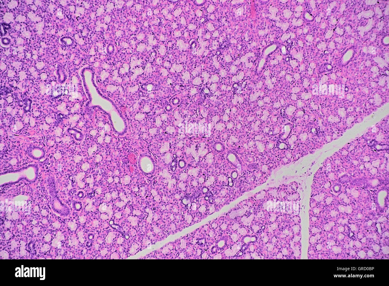
Histology, Salivary Glands Biology Diagrams
The salivary glands are exocrine glands that make, modify and secrete saliva into the oral cavity. Human salivary glands produce between 0.5 to 1.5 L of saliva daily, facilitating mastication, swallowing, and speech, lubricating the oral mucosa, and providing an aqueous medium for taste perception. The salivary glands: anatomy and The minor salivary glands secrete saliva continuously, keeping the mouth optimally moist. When food enters, the major glands activate and large amounts of saliva pour out. The average human being produces around 1500ml of saliva per day, but it can be a great deal higher if the glands are stimulated properly.