SOLUTION Spleen and its functions Biology Diagrams Discover the anatomy of the spleen, including its location, borders, surfaces, extremities, capsule, and ligaments. Explore in depth its histology, neurovascular supply and functions. Functions of spleen. The spleen has several functions. While it is an essential organ, it is not a vital one. A person can live everyday life without a spleen Structure and Anatomy. The spleen is a soft, highly vascular, fist-sized organ that plays a vital role in the lymphatic and circulatory systems. It is encased in a fibrous capsule and consists of two main types of tissue—red pulp and white pulp—which perform distinct functions related to blood filtration and immune response. Below is a

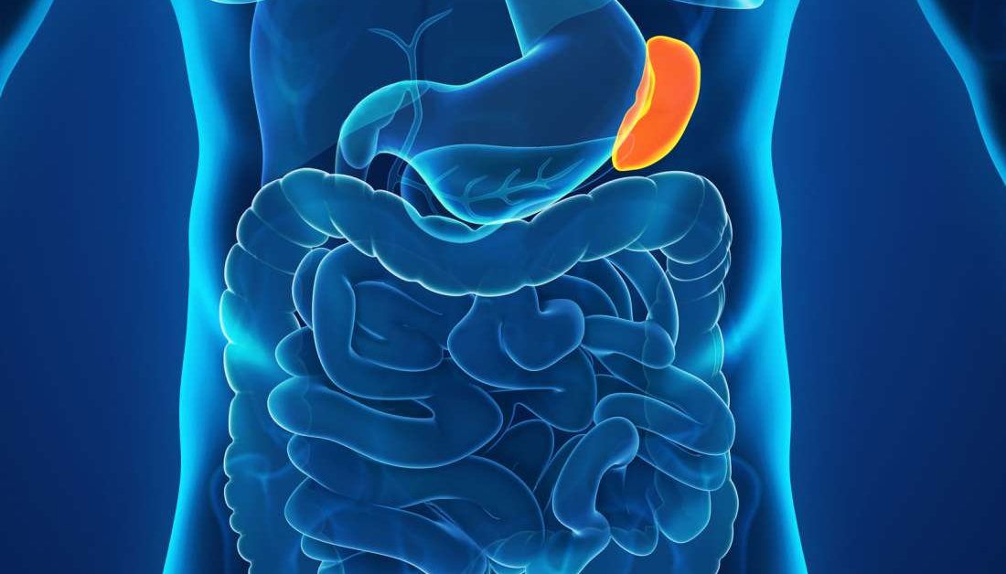
The spleen is a significant organ of the hematologic and reticuloendothelial systems. It is an intraperitoneal organ located in the left upper quadrant of the abdomen posterior and lateral to the stomach.[1] The spleen is situated anatomically behind the 9 and 11 ribs on the left side of the body. The spleen is a small organ at the top of the abdomen. It filters the blood and helps defend the body against pathogens. In this article, we will explain its anatomy, what it does, and what
Anatomy, Abdomen and Pelvis, Spleen Biology Diagrams
The Anatomy of the Spleen The spleen sits in the upper left abdomen just below the diaphragm. By Mark Gurarie. Updated on December 10, 2024. Medically reviewed by Robert Burakoff, MD. Spleen function can become hampered by sickle-cell anemia, a disease in which the shape of RBCs is affected. Spleen Anatomy. A dense connective tissue capsule covers the spleen. Trabeculae extend inward, dividing the organ into lobules. Invertebrates do not have a spleen. Functions of the Spleen. The spleen performs multiple critical functions: Hematopoiesis: The spleen produces blood cells in a fetus. While it stops producing red blood cells
spleen, organ of the lymphatic system located in the left side of the abdominal cavity under the diaphragm, the muscular partition between the abdomen and the chest. In humans it is about the size of a fist and is well supplied with blood.As the lymph nodes are filters for the lymphatic circulation, the spleen is the primary filtering element for the blood. Microscopic anatomy. Understanding the microscopic anatomy of the spleen is important for understanding its function. Numerous septa called trabeculae extend from the dense irregular fibroelastic connective tissue of the capsule into the parenchyma of the spleen. Both the capsule and trabeculae contain myoepithelial cells which have the ability to contract.
